Mammograms – screening and clarifying changes in the breast
What is a mammogram?
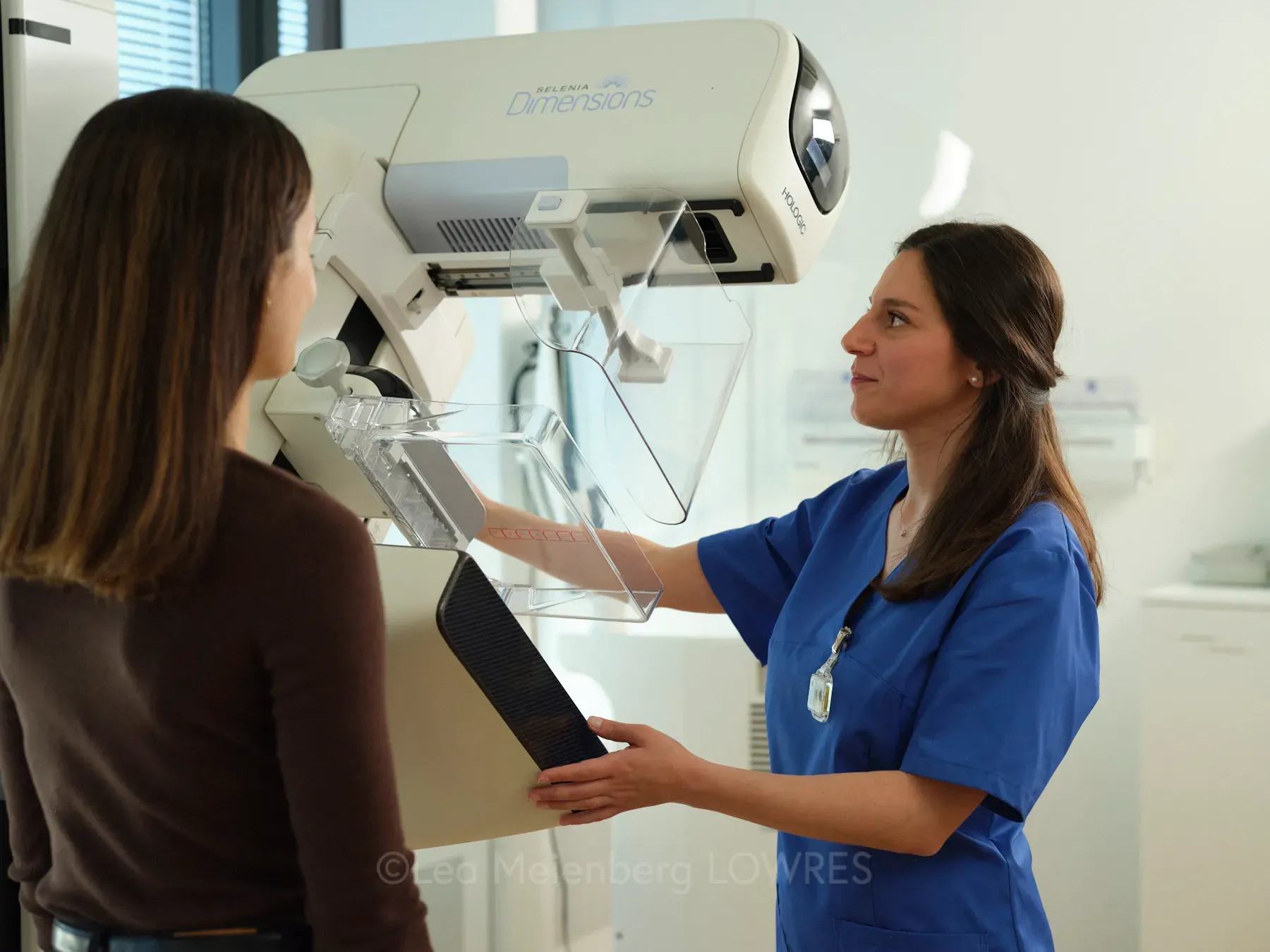
A mammogram is a special X-ray examination that can render even the smallest changes in breast tissue visible. It often reveals microcalcifications, dense tissue and early stages of tumours even before symptoms appear. Brief, controlled compression of the breast produces clear, structured images, enabling our radiologists to deliver a reliable assessment of the glandular tissue. The radiation dose used is low and is considered medically well tolerated. Cutting-edge digital systems allow for images to be saved and accurately compared with previous images so that even minimal changes in tissue can be detected.
Why and when is a mammogram needed?
Mammograms are used both for screening purposes and for clarifying specific symptoms in the breast. In many regions, women aged 50 and over are regularly invited to participate in screening programmes in order to detect changes at an early stage. The examination may also be useful for younger women or patients with a heightened family risk.
Typical reasons for a mammogram are:
- New palpable lumps or hardenings
- Abnormal findings on an ultrasound
- Unexplained pain in breast
- Family history or genetic risk
- Follow-up checks after breast surgery or tumour treatment
- Early detection (screening)
In cases of dense breast tissue, a mammogram is often undertaken in addition to ultrasound scans or magnetic resonance imaging to provide a full diagnostic picture.

How do I prepare for the examination and what does it involve?
Preparation is straightforward. If you experience breast pain, the examination should take place in the first week after your period, as this is when the breast tissue is less sensitive. On the day of the examination, you should refrain from using deodorants, creams or powders in the breast and armpit area, as residues can impair the image quality.
During the examination, you will be accompanied by experienced specialists. Each breast is positioned one at a time, then compressed and images are taken in several planes. This compression may cause brief discomfort, but it is necessary to achieve optimal image quality and reduce the radiation dose. The examination itself takes only a few minutes in total. The images are then checked for technical quality to ensure that all relevant areas are as clearly visible as possible.
Risks, limitations and complementary procedures
Mammograms are very safe, and involve only low radiation exposure. Nevertheless, as with any medical examination, not every finding can be conclusively assessed. Particularly dense breast tissue can make it difficult to interpret images, making additional procedures necessary.
Where necessary, the following methods are used:
- Breast ultrasound for additional imaging of glandular and connective tissue
- Magnetic resonance imaging (MRI) in cases of heightened risk or unexplained findings
- Tissue sample (biopsy) for further diagnosis if imaging findings are inconclusive
This combination of different methods enables a very high degree of diagnostic certainty.
Aftercare, findings and regular check-ups
Once the examination is complete, the images are evaluated by our specialist consultants. The findings are explained in writing or in a personal consultation. If the results are normal, a repeat mammogram is usually recommended after approximately two years as part of the screening programme.
For women with a heightened risk, dense breast tissue or a history of breast cancer, shorter check-up intervals may be advisable. Digital diagnostic systems enable specialists to make detailed comparisons of progression, so that changes can be detected at an early stage.
Cutting-edge technologies in breast diagnostics
In addition to conventional mammograms, innovative imaging techniques are available today. These include tomosynthesis, which produces layered images of the breast tissue and avoids overlaps. This technology improves the visualisation of small tumours, particularly in dense breast tissue.
Contrast-enhanced mammography
We also offer contrast-enhanced mammograms, which can provide more information about the breast – comparable to an MRI. With this method, an iodine-based contrast medium is injected into a vein before a special mammogram is performed. This method can reveal changes that are not yet detectable using other standard imaging procedures.